
Scene Preview
study:
Adjacent domain-general and sensory-biased regions
SCENE FILE:
MD_modality_ms_rev
SCENE:
Figure_1
DESCRIPTION:
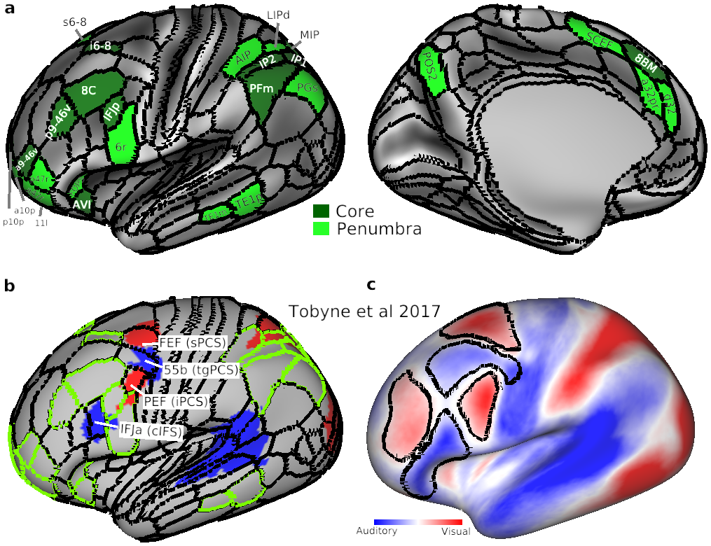
(a) The extended MD system. Core MD regions are colored in dark green and white labels. Penumbra MD regions are in light green with black labels. Note here we separated core region SCEF/8BM (as identified in Assem et al 2020) into SCEF as penumbra and 8BM as core for simplicity in analysis. (b) and (c) are adapted from Tobyne et al. (2017). (b) Sensory-biased regions [originally identified in (Michalka et al. 2015)] after their transformation to the HCP fs_LR surface. Red: visually biased. Blue: auditory biased. Overlapping HCP MMP1.0 regions are labelled and their original Michalka et al. (2015) labels are in brackets. Green contours correspond to extended MD borders in (a). (c) Sensory-biased lateral frontal regions based on their intrinsic rfMRI connectivity with posterior cortical areas. Black contours surrounding regions with warmer colours (red) are significantly more connected with visual parietal areas than auditory temporal regions. Black contours surrounding regions with colder colours (blue) are significantly more connected with auditory temporal regions than visual parietal regions
TAGS:
Species:Human